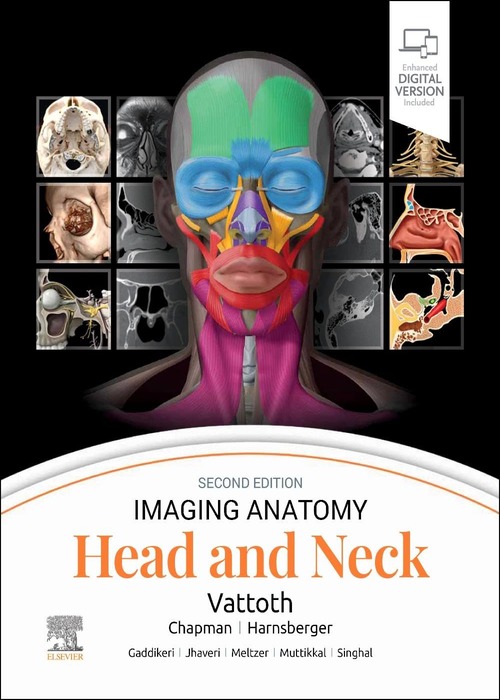
Imaging Anatomy: Head and Neck: 2ed
This richly illustrated and superbly organized text/atlas is an excellent point-of-care resource for practitioners at all levels of experience and training.
Not in Stock. Ships in: 4-6 weeks
Details
Imaging Anatomy: Head and Neck: 2ed
This richly illustrated and superbly organized text/atlas is an excellent point-of-care resource for practitioners at all levels of experience and training. Written by global leaders in the field, Imaging Anatomy: Head and Neck, second edition, provides a thorough understanding of the detailed normal anatomy that underlies contemporary imaging. This must-have reference employs a templated, highly formatted design; concise, bulleted text; and state-of- the-art images throughout that identify the clinical entities in each anatomic area, offering a unique opportunity to master the fundamentals of normal anatomy and accurately and efficiently recognize pathologic conditions.
Key Features
- Features hundreds of detailed, full-color illustrations and more than 900 high-resolution, cross-sectional radiologic images that together illustrate the fine points of imaging anatomy for new and experienced head and neck imaging specialists
- Contains new chapters on external nose anatomy, the facial nerve in temporal bone, minor fissures and sutures around the temporal bone, and temporal bone anatomy on photon-counting detector (PCD) CT
- Provides updated, enlarged images and captions in areas such as facial muscles and the superficial musculoaponeurotic system, and frontal recess and related air cells
- Includes extensive new content on PCD CT; new details on relatively unknown anatomical foramina, such as the vomerovaginal canal and canaliculus innominatus; new content based on the International Frontal Sinus Anatomy Classification; and minute details on the course of nerves in the head and neck
- Includes a series of successive imaging slices in each standard plane of imaging (coronal, sagittal, and axial) to provide multiple views that further support learning
- Depicts common anatomic variants and covers the common pathological processes that manifest with alterations of normal anatomic landmarks
- Reflects new understandings of anatomy due to ongoing anatomic research as well as new, advanced imaging techniques
- Presents essential text in an easy-to-digest, bulleted format, enabling imaging specialists to find quick answers to anatomy questions encountered in daily practice
| Book | |
|---|---|
| Author | Vattoth, Surjith |
| Pages | 664 |
| Year | 2024 |
| ISBN | 9780443249648 |
| Publisher | Elsevier |
| Language | English |
| Uncategorized | |
| Edition | 2/e |
| Weight | 1.81 kg |
| Dimensions | 21.59 x 27.94 x 3.81 cm |
| Binding | Hardcover |
| Imprint | Elsevier |


